Best Pulmonology Hospital with Expert Lung Care Services

From respiratory disorders to lung health optimization, we’re here to help you breathe easier and live better. Our dedicated team of pulmonologists combines advanced diagnostics with compassionate care to address a wide range of lung conditions. To care for your lung health visit our skilled pulmonologists.

It is no exaggeration to say that the Department of Pulmonology of Worlds Nic is among the foremost of its kind in the country. Our department comprises the best pulmonologists. The equipment, facilities, and expertise in diagnosing and treating diseases of the respiratory tract have been set to global benchmarks. We were the first in India to have an exercise physiology test system with breath-to-breath analysis of respiratory gases.
Our Lungs Specialist routinely performs bronchoscopies, thoracoscopies, pleural drainage procedures, pleurodesis, CT-guided FNAC (fine needle aspiration cytology), and FNAB (fine needle aspiration biopsy).
Patients suffering from hemoptysis benefit from bronchial artery embolization while those affected by diffuse lung disease benefit from cryo-biopsies. If you suffer from pulmonary disorders, our lungs doctor can help you.

Medical Conditions

Asthma is a chronic inflammatory illness of airways that can affect anyone at any age. As a result of inflammation and muscle tension around the small airways, the airways in the lungs become congested.

Shortness of breath and chest tightness are all symptoms of asthma that can be caused by this. It is common for these symptoms to be worse at night or during physical activity because they are transient in nature.

Asthma symptoms might be aggravated by a variety of other typical factors. Viral infections (colds), dust, smoke, fumes, changes in temperature, grass and tree pollen, animal hair and feathers, strong soaps and perfumes are just a few examples of what can set off an allergic reaction in different people. The best pulmonologist helps you find the right treatment for asthma.

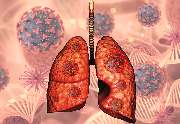
An illness called pneumonia causes the air sacs in one or both lungs to become inflamed. Coughing up phlegm or pus, fever, chills, and difficulty breathing are all major lung infection symptoms that can result from the air sacs being swollen with fluid or pus (purulent material). One of the most common causes of pneumonia is caused by a range of microorganisms.

Pneumonia can be minor or life-threatening, depending on the severity of the infection. Infants and small children, the elderly, and those with health issues or compromised immune systems are at greatest risk. Expert Pulmonologists Worlds Nic offers state-of-the-art facilities that help in the treatment of pneumonia.

Edema of the lungs' tissues and air spaces (typically alveoli) is referred to as "pulmonary congestion'' when there is an excessive buildup of liquid in the tissues and spaces. As a result, hypoxemia and respiratory failure are possible outcomes.

The major cause of pulmonary edema is when the left ventricle fails.

When lung tissue blood vessels are damaged (non-cardiogenic pulmonary edema). The three main goals of treatment are to improve respiratory function, address the underlying cause, and avoid additional damage to the lungs. Respiratory failure or cardiac arrest can occur as a result of hypoxia in pulmonary edema develops suddenly (acutely). The best pulmonologists at Worlds Nic strive to provide advanced facilities to treat respiratory related concerns.

Pulmonary embolism (PE) is the obstruction of a lung artery by a material that has traveled via the bloodstream from another part of the body (embolism). PE symptoms include shortness of breath, chest pain, especially while inhaling, and coughing up blood.

There may also be signs of a blood clot in the leg, such as a red, heated, swollen, and painful leg. Low blood oxygen levels, fast breathing, high heart rate, are symptoms of pulmonary embolism.

The diagnosis of pulmonary embolism may be confirmed by CT pulmonary angiography, lung ventilation/perfusion scan, or ultrasound of the legs. DVT and PE are often referred to as venous thromboembolism (VTE).

Due to abnormalities in the tiny airways of the lungs, airflow in and out of the lungs is restricted. Multiple processes contribute to the constriction of the airways. Parts of the lung may be destroyed, mucus may obstruct the airways, and there may be inflammation and edema of the airway lining.

COPD has emphysema or chronic bronchitis components. Typically, emphysema refers to the destruction of the tiny air sacs at the end of the lung airways. Chronic bronchitis refers to a persistent cough accompanied by phlegm production caused by inflammation in the airways.

COPD and asthma share similar symptoms (cough, wheezing, and breathing trouble), and individuals can have both illnesses. Here at Worlds Nic, our expert pulmonologists have had success treating chronic obstructive pulmonary disease.

Treatments & Procedures

This is a procedure by which a bronchoscopist collects specimens from the distal most airways. Aliquots Saline is instilled into the affected segment via the working channel of bronchoscope then suctioned back. The retrieved fluid is then sent for various microbiological analysis & cytology. This is an excellent diagnostic tool for cancer and infections. This procedure is performed by expert pulmonologists to ensure you receive a safe treatment.

This is a procedure by which a bronchioscopist collects specimens from the distal most airways. Aliquots Saline is instilled into the affected segment via the working channel of bronchoscope then suctioned back. The retrieved fluid is then sent for various microbiological analysis & cytology. This is an excellent diagnostic tool for cancer and infections.

Worlds Nic routinely uses this procedure for histopathological diagnosis of diffuse parenchymal lung disease. In this transbronchial lung biopsy, forceps are passed through the working channel in the bronchoscope at the point of concern with the help of biopsy forceps. Biopsy of affected lung tissue is done. The biopsy sample is then sent for pathological examination.

In certain cases, the specimens obtained in a transbronchial lung biopsy are inadequate or insufficient to diagnose certain conditions. The specimens which are obtained by the forceps are too small for diagnosis of diffuse lung disease. However, the latest technology has provided us with a far more effective new option. Worlds Nic offers its patients the advantages of cryobiopsy. A flexible ‘cyroprobe’ is guided to the location from which a specimen is required. It is then cooled using a variety of technologies. This cooling freezes the tissue of the lungs in the immediate region and it adheres to the tip of the probe, after which it is retracted. The size of the specimen obtained by this method is consistently larger than in the traditional way and accuracy of diagnosis has been proven to be much higher.

Endobronchial Ultrasound (EBUS) guided Transbronchial Needle Aspiration (TBNA)

Known as EBUS TBNA for short, this is a specialized technique used to obtain body tissue samples from Thoracic Lymph Nodes. It uses a special type of bronchoscope, which has ultrasound at its tip. In conventional TBNA, the procedure is done without guidance of Ultrasound. In EBUS-guided TBNA, the procedure is guided by Ultrasound, hence diagnostic accuracy and yield are much better.

A thoracoscopy is a minimally invasive procedure used for both diagnostic and therapeutic purposes. A thoracoscope (a thin, flexible tube with a camera) is inserted into the Pleural Cavity via an incision so that both visceral and parietal pleura (a membrane covering the lungs) are examined.

It conducted for the following conditions:

Pleural Biopsies for diagnosis of mesothelioma and malignant pleural effusion

To remove excess air or fluid from the region surrounding the lungs

To remove pleural cysts or diseased lung tissue

To lyse the adhesion in the pleural cavity secondary to complicated parapneumonic effusion and

Pleurodesis – Procedure by which Talc is insufflated into the Pleural cavity to prevent recurrence of malignant pleural effusion (accumulation of liquid in the pleural cavity secondary to malignancy)

Pneumothorax (accumulation of air in the pleural cavity)

When a person coughs up blood it is known as haemoptysis (or hemoptysis). This can be due to various causes like infection, cancer, aneurysm (dilation & thinning of blood vessel) and bronchiectusis (dilation & destruction of bronchus). Haemoptysis is a medical emergency and requires immediate attention, evaluation and treatment. Bronchial artery embolisation is one of the more advanced techniques practiced at Sri Ramakrishna Hospital to stop hemoptysis. It is a minimally invasive procedure. The doctor inserts a catheter into an artery in the leg, which is then guided up to the lungs. A dye is injected to identify the source of the bleeding. The affected artery is then blocked through the use of metal coils or other techniques and substances. This stops the bleeding. The blocked artery is not a concern since other arteries immediately compensate for it.

FNAC and FNAB are procedures wherein cells or tissue are removed from the body for pathological examination. Lung biopsies can now be performed using a minimally invasive technique under CT guidance. A fine needle with a hollow end is inserted into the lung mass / nodule and Tissue obtained. This can be done without a patient having to be on general anaesthesia. The aspirate (FNAC) is made into smears for cytological evaluation and the tissue (biopsy specimen) taken is subjected for pathological examination.

GET ANSWERS

Frequently Asked Questions

Lung infections are quite common and can occur due to pollution or other factors. Few major signs that can help identify a lung infection include, Troubled breathing Continuously gasping for air Continuous cough Upper abdominal pain while breathing
Noisy breathing or wheezing indicates that something unexpected is clogging or narrowing your lungs' airways. Coughing up blood could indicate a problem with your lungs or upper respiratory system. It's a symptom of a health condition, regardless of where it's coming from.
Irritants that damage your lungs and airways over time create chronic obstructive pulmonary disease (COPD), which includes chronic bronchitis and emphysema. COPD is primarily caused by smoking, but it can also affect nonsmokers.
Sharp or stabbing chest discomfort is common when a lung infection is present. When coughing or breathing deeply, the chest pain tends to get worse. Sharp sensations might sometimes be felt in your mid to upper back.
When people with COPD have flare-ups, their symptoms get a lot worse, and they may need extra care at home or to go to the clinic for emergency care. Severe flare-ups can put a person's life in danger.
Most lung infections should get better in a few weeks with rest, care to make the person feel better, and sometimes antibiotics. But if your signs last longer than this, you might have a long-term lung infection.